
1. Image guided neuro-intervention
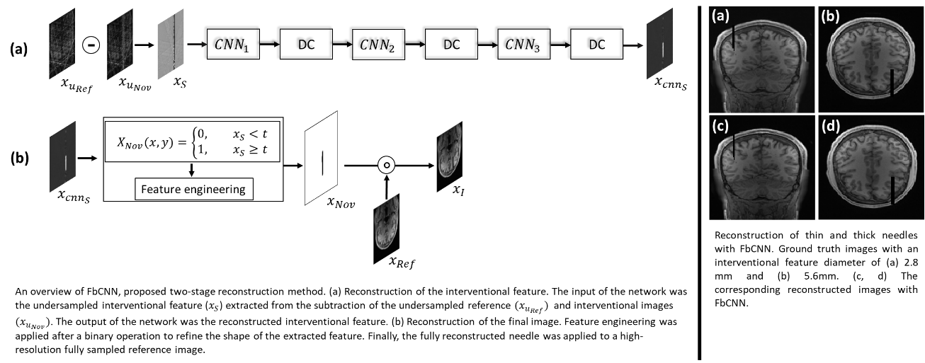
Feature-based convolutional neural network (FbCNN) for reconstruction of interventional MRI
Interventional MRI (I-MRI) provides exceptional advantages to other imaging modalities in image-guided neurosurgery. Our aim is to achieve high spatial and temporal resolution I-MRI for neuro-intervention. In terms of image acquisition and reconstruction, we proposed a novel feature based image reconstruction scheme using golden-angle sampling and machine learning.
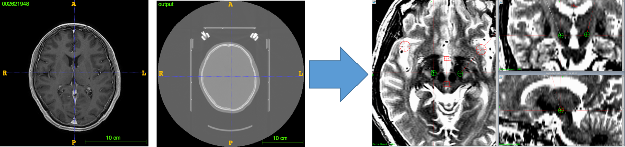
Machine-learning based post-operative electrode placement prediction in deep brain stimulation
Deep brain stimulation (DBS) is a neurosurgical procedure for treating neurodegenerative diseases and neurological disorders such as Parkinson’s disease (PD) and epilepsy. Image guidance is crucial for the accurate placement of DBS electrodes. However, current surgical planning systems based on pre-operative MR and CT images of the brain cannot take into account the intra-operative brain-shift, resulting in suboptimal electrode placement undermining clinical outcomes. In this study, a support vector regression (SVR) model was constructed based on 114 patient-specific data of PD patients. Two target nuclei were manually delineated based on pre-operative MR and CT images. Spatial coordinates of the two nuclei were collected and compared to the post-surgical electrode position from CT images. Analysis of a total of 45 features showed that the pre-operative target coordinates are the parameters mainly influencing the model prediction for both nuclei. The mean absolute error (MAE) of the prediction of the electrodes on unseen patients was 0.76mm. This study demonstrates the potential of using SVR modelling to improve current DBS surgical planning procedure and pre-operative risk-assessment.
Deep brain stimulation (DBS) is a neurosurgical procedure for treating neurodegenerative diseases and neurological disorders such as Parkinson’s disease (PD) and epilepsy. Image guidance is crucial for the accurate placement of DBS electrodes. However, current surgical planning systems based on pre-operative MR and CT images of the brain cannot take into account the intra-operative brain-shift, resulting in suboptimal electrode placement undermining clinical outcomes. In this study, a support vector regression (SVR) model was constructed based on 114 patient-specific data of PD patients. Two target nuclei were manually delineated based on pre-operative MR and CT images. Spatial coordinates of the two nuclei were collected and compared to the post-surgical electrode position from CT images. Analysis of a total of 45 features showed that the pre-operative target coordinates are the parameters mainly influencing the model prediction for both nuclei. The mean absolute error (MAE) of the prediction of the electrodes on unseen patients was 0.76mm. This study demonstrates the potential of using SVR modelling to improve current DBS surgical planning procedure and pre-operative risk-assessment.

2. Brain modeling
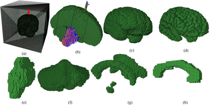
We aim to construct a biophysically accurate brain model for various applications, such as traumatic brain injury (TBI), brain intervention, and brain development. The biomechanical properties of the brain were measured by ex vivo mechanical testing and in vivo MR elastography imaging. The boundary conditions of the brain were acquired by in vivo motion imaging techniques. Based on the brain properties and models, we study the mechanism of TBI, brain deformation during intervention, and the brain development patterns and related diseases.
We aim to construct a biophysically accurate brain model for various applications, such as traumatic brain injury (TBI), brain intervention, and brain development. The biomechanical properties of the brain were measured by ex vivo mechanical testing and in vivo MR elastography imaging. The boundary conditions of the brain were acquired by in vivo motion imaging techniques. Based on the brain properties and models, we study the mechanism of TBI, brain deformation during intervention, and the brain development patterns and related diseases.
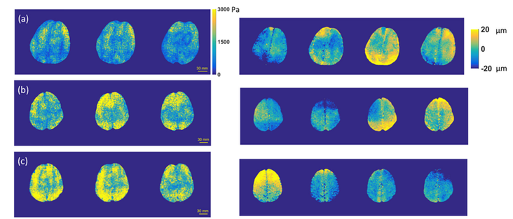
Estimated shear moduli and wave images of the brain from a healthy volunteer at actuation frequencies of (a) 50 Hz, (b) 65 Hz, and (c) 80 Hz. The displacement component was shown in the z (foot-head) direction
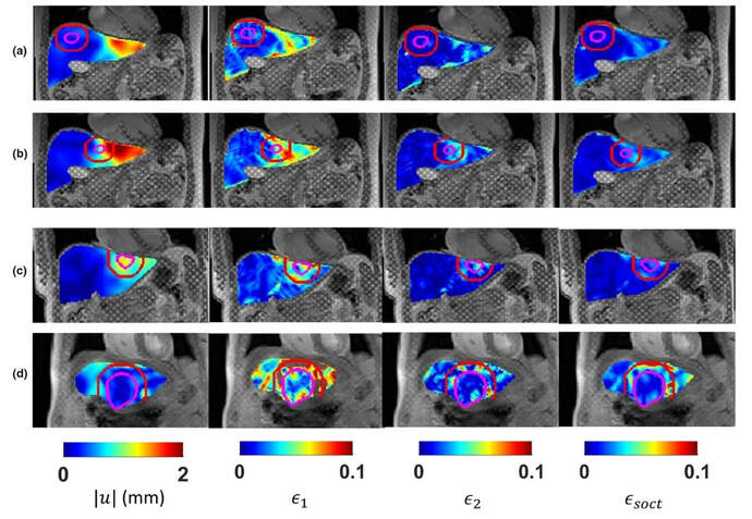
MR image-based brain interventional modeling
MR tagging based tumor detection
